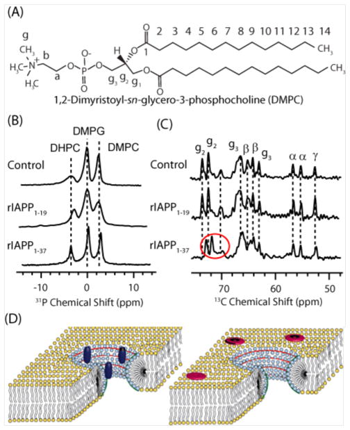
Figure 4. Toxic variants of IAPP.
(A) 31P NMR spectra showing binding of toxic rIAPP1–19 to DHPC in bicelles. (B) 13C NMR spectra showing binding of non-toxic rIAPP1–37 to DMPC and DMPG. (C) Cartoons of bicelles showing rIAPP1–19 (dark blue) localized to DHPC in the curved perforation (light blue) and rIAPP1–37 (red) localized to DMPC and DMPG in the flat lamellar region (yellow). Adapted from reference 19.

