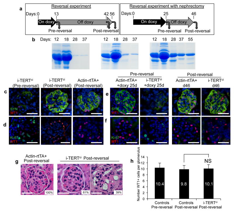
Figure 5.
Proliferating podocytes return to a quiescent, differentiated state after doxycycline withdrawal in i-TERTci mice. (a) For TERT reversal experiment (left), 6 i-TERTci mice were treated with doxy, three mice were sacrificed at day 13 (Pre-reversal) and doxy was removed from the remaining three mice, which were aged until at least 42 days (Post-reversal). For reversal with nephrectomy (right), two Actin-rtTA+ and two i-TERTci mice were treated with doxycycline. Each mouse underwent survival nephrectomy at day 25, doxy was withdrawn after surgery and the mice were aged until day 46. (b–d) Reversal experiment. (b) Serial analysis of protein in urine by SDS-PAGE in three i-TERTci mice treated with doxy as described in (a, left). (c, d) Double immunostaining for synaptopodin (SYN, green) and WT1 (red) (c) or for Ki-67 (green) and WT1 (red) (d) in kidney sections from i-TERTci mice on doxy for 13 days (+ doxy), and i-TERTci or Actin-rtTA+ mice on doxy for 13 days then reversed for 29 days. Scale bar = 25 μm. (e–g) Reversal experiment with nephrectomy. (e,f) Double immunostaining for synaptopodin (SYN, green) and WT1 (red) (e) or for Ki-67 (green) and WT1 (red) (f) in kidney sections from nephrectomized i-TERTci and Actin-rtTA+ control mice, as described in (a, right). Scale bar = 25 μm. (g) H&E stained sections of post-reversal kidney from nephrectomized i-TERTci or Actin-rtTA+ mice at day 46. Percentage of glomeruli showing a morphology similar to that outlined is indicated. Scale bar = 25 μm. (h) Quantification of podocyte density in glomeruli of control mice pre- and post-reversal, and in glomeruli of i-TERTci mice after reversal. NS, not significant. P=0.39, t-test for i-TERTci post-reversal vs. controls post-reversal.

