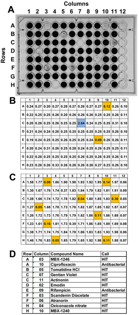
Figure 5.
An example of the screening results obtained using S. epidermidis 18972 and a mock screening plate. A) A photograph of the crystal violet stained mock screening plate that contained nine compounds that are inhibitors of biofilm formation and two antibiotics. The remainder of the wells in columns 2–11 contained DMSO. Columns 1 and 12 contain the 0% INH (DMSO only) and 100% INH (100 µg chloramphenicol/ml; Cm100) controls, respectively. The wells containing anti-biofilm and antibacterial compounds exhibited significant reductions in the intensity of crystal violet staining. B) The OD600 values obtained for the planktonic cultures. Compounds that produced ≥ 40% inhibition planktonic growth are highlighted in orange. The compound in well number C07, highlighted in blue, produced an OD600 of 2.64, which is significantly higher than those produced by the 0% INH (DMSO only) controls. This well contained gentian violet, an intensely colored compound that absorbs light at 600 nm. Beware of colored compounds that absorb at 600 nm, as they can mask antibacterial activity. C) The OD600 values obtained for the crystal violet stained biofilm cultures. Compounds that produced ≥ 80% inhibition planktonic growth are highlighted in orange. D) A table listing the compounds used in the mock screening plate, their location on the plate (row, column), and the screening call (HIT or antibacterial). A compound that produced % ≥80% biofilm inhibition and ≤ 40% planktonic growth inhibition were designated anti-biofilm “HITS”. Compounds that produced ≥ 40% planktonic growth inhibition were designated as “Antibacterial” compounds. Seven of the antibiofilm compounds used in the mock screening plate were identified previously in a pilot screen using a compound library comprised of 2000 known bioactive chemicals (Spectrum library; MicroSource Discovery Systems, Gaylordsville, CT). MBX-1240 and MBX-1246 were identified in screen of compounds purchased from Chembridge Corp. (San Diego, CA) (Opperman et al., 2009)

