
Figure 3.
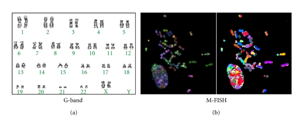
Karyotype of cultured human PDL cells. (a) G-banding staining revealed that the number, banding, and shape of the chromosomes were normal. (b) Multiplex fluorescence in situ hybridization (M-FISH) that visualises each chromosome in a different colour showed that no chromosomal aberrations, including translocation, had occurred.