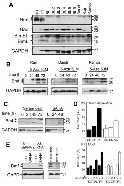
Figure 7. Bmf expression is absent in Burkitt lymphoma cells but can be restored upon demethylation.
(A) Western blot analysis of Bmf, Bim and Bad in CD19+ cells derived from peripheral blood of healthy donors (#1 and #2), primary Burkitt lymphoma samples (BL1-BL6) and BL cell lines Daudi, Raji and Ramos. (B) Western blot analysis of Bmf levels in BL cell lines after inhibition of DNA-methyltransferases with 5-aza-2′-deoxycytidine (5μM) for the indicated times. (C) Western blot analysis of Bmf levels in Ramos cells after serum deprivation or inhibition of with SAHA (2μM) for the indicated times. (D) Cell death determined by AnnexinV/7-AAD staining in Ramos cells expressing either an shRNA against Bmf (white bars) or an unspecific shRNA (black bars) after serum deprivation or treatment with 2 or 4μM SAHA for the indicated times. Values represent mean±SE of 3 independent experiments. (E) Efficiency of knock down was confirmed by Western blot analysis of Bmf levels in cells treated with 2μM SAHA for 48 hours (left panel) or cells deprived of serum for 48 hours (right panel).

