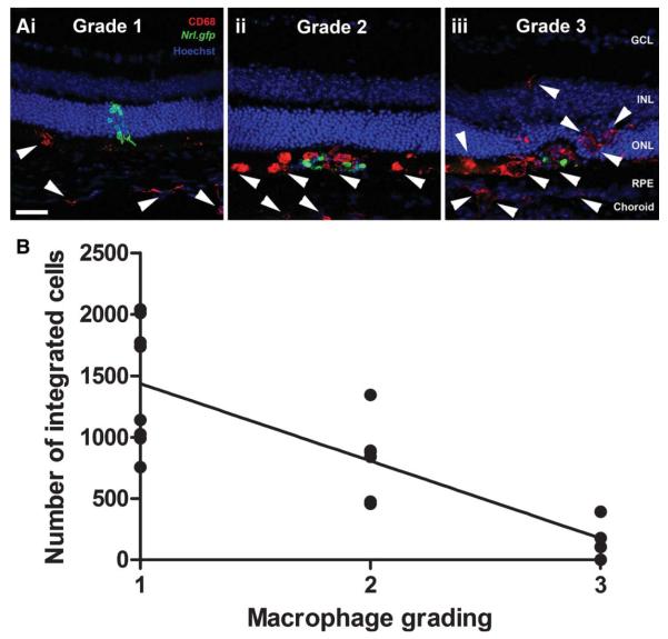
Figure 2.
Assessment of macrophage activation and photoreceptor cell integration. (A): Projection confocal images showing the grading scheme used to assess macrophage activation and infiltration in transplanted eyes (CD68, an activated macrophage marker, red; white arrow heads). (B): Line graph showing a negative correlation between the number of integrated cells and level of macrophage activation (Pearson correlation, p < .0001, n = 18). Scale bar = 50 μm. Abbreviations: GCL, ganglion cell layer; INL, inner nuclear layer; ONL, outer nuclear layer; RPE, retinal pigment epithelium.

