Figure 1.
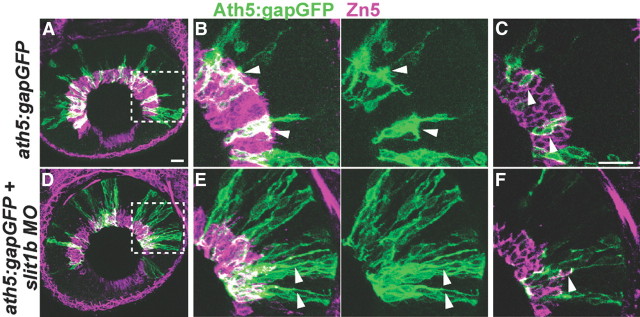
Apical processes retraction of RGCs is delayed in slit1b morphants. Analysis was performed on ath5:gapGFP DNA-injected embryos at 48 hpf. A, D, Extended-focus confocal images of the retinas of a control embryo (A) or a slit1b morphant (D). Boxed regions in A and D are enlarged in B and E, respectively. Arrowheads point to the retracted apical processes in the control retina (B) and un-retracted apical processes in the slit1b morphant retina (E). Zn5 (magenta) labels the ganglion cell soma and axon. C, F, Single optical section images of the boxed regions in A and D, respectively, showing that the Ath5:gapGFP+ cells express Zn5 and are thus RGCs (arrowheads). Scale bars, 20 μm.

