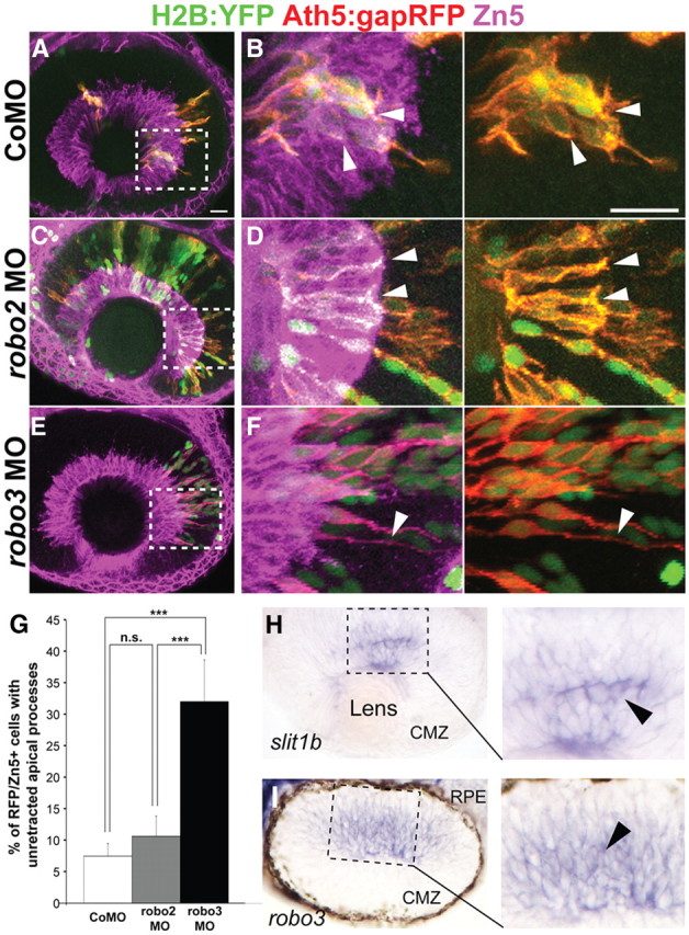
Figure 2.

Knockdown of robo3 but not robo2 inhibits apical process retraction. A–F, Confocal images from wild-type hosts transplanted with blastomeres from Tg(ath5:gapRFP) embryos injected with control morpholino (CoMO; A, B), robo2 morpholino (C, D), or a combination of robo3v1 and v2 morpholinos (E, F) at 48 hpf. Zn5 (magenta) labels the ganglion cell layer. Boxed regions in A, C, and E are enlarged in B, D, and F, respectively. Arrowheads in B and D indicate the retracted apical processes of transplanted Ath5:gapRFP+ RGCs from control or robo2 morpholinos-injected embryos; arrowheads in F indicate the un-retracted apical process of a transplanted RGC from a robo3v1 and v2 double-morphant. Scale bars, 20 μm. G, Percentage of Ath5:gapRFP and Zn5 double-positive cells with un-retracted apical processes at 48 hpf (n = 187 cells from control morpholino-injected embryos; n = 94 cells from robo2 morphants; n = 50 cells from robo3 morphants). Error bars indicate SEs (***p < 0.001). H, I, A retinal section from a 38 hpf whole-mount in situ hybridized embryo, showing that slit1b and robo3 mRNAs are expressed in the developing zebrafish retina. I, In situ hybridization image from a similarly staged retinal section showing that robo3 mRNA is expressed in subsets of cells in the developing retina. Note that pigments were allowed to form and the retinal pigmented epithelium (RPE) is thus visible. CMZ, Ciliary marginal zone.
