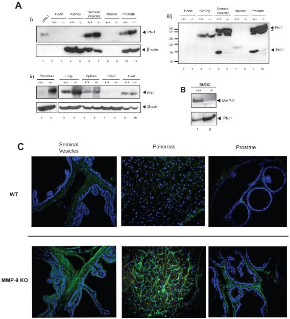
Figure 2. PN-1 is targeted by MMP-9 in vivo.
A) Tissue lysates of indicated organs from C57/B6 WT (+/+) or MMP-9 KO (−/−) mice were resolved by SDS-PAGE gel and blotted with anti-mouse PN-1 or anti-β-actin antibody (i and ii). 20ng recombinant mouse PN-1 protein (rPN-1) was a control. Please note β-actin is not present in heart or muscle. Panel iii) shows Panel i) after longer exposure. A PN-1 degradation fragment was now apparent in the lysates from the seminal vesicles and the prostate. B) Primary bone marrow derived cells (BMDC) were grown from C57/B6 WT (+/+) or MMP-9 KO (−/−) mice, serum free conditioned media was subjected to immunoblot with anti-MMP-9 or anti-PN-1 antibody. C) Immunohistochemistry showing the expression of PN-1 (green) in seminal vesicles, pancreas and prostate from WT or MMP-9 KO mice. Cell nuclei were stained by Hoechst (blue).

