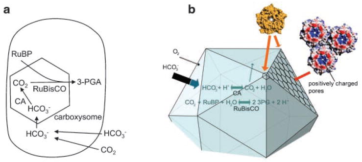
Figure 2.
The bacterial carbon dioxide concentrating mechanism. a: The bacterial CCM starts with transport of inorganic carbon into the cell as HCO3−. Equilibrium between HCO3− and CO2 is not reached due to a lack of carbonic anhydrase in the cytoplasm. HCO3− is converted to CO2 by carboxysomal carbonic anhydrase within the microcompartment lumen. The protein shell of the carboxysome impedes CO2 diffusion. Consequently, CO2 can accumulate in the immediate vicinity of RuBisCO while diffusive loss of CO2 through the cell membrane is minimized. Elevated CO2 proximal to RuBisCO increases carbon fixation and suppresses photorespiration (a nonproductive process in which O2 replaces CO2 as a substrate for RuBisCO). b: A preliminary atomic model of the carboxysome shell based on crystal structures of the component shell proteins.(12) The positively charged pores through the sheet of hexagonal proteins are indicated. These have been postulated to enhance the diffusion of negatively charged molecules such as bicarbonate (thick arrow) across the shell, compared to uncharged molecules such as CO2 and O2 (thin arrows).

