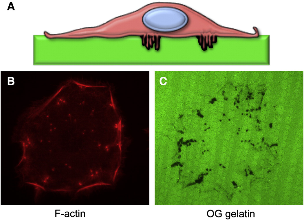
Figure 2. Invadopodia are invasive microdomains.
- Diagram of a cancer cell growing on top of a fluorescent ECM substrate. Invadopodia are thin cellular protrusions from the cell-substrate interface into the substrate.
- F-actin staining of a head and neck carcinoma cell showing invadopodia (dots).
- OregonGreen-labeled gelatin from the same field as B) showing the areas of invadopodia degradation (black dots).

