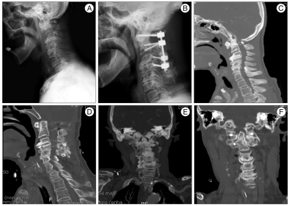
Fig. 2.
Case 2 image findings. A : Preoperative X-ray showing severe kyphosis with degenerative spondylotic change and increased ADI about 9 mm and BI. B : Postoperative X-ray showing correction of cervical kyphosis and reduction of BI as well as C1-4 fixation. C-F : Compared with preoperative CT, postoperative CT showing decompressed craniovertebral junction and distracted joint space in which in which autograft iliac bone spacer (arrow) have been placed. ADI : atlanto-dental interval, BI : basilar invagination, CT : computed tomography.

