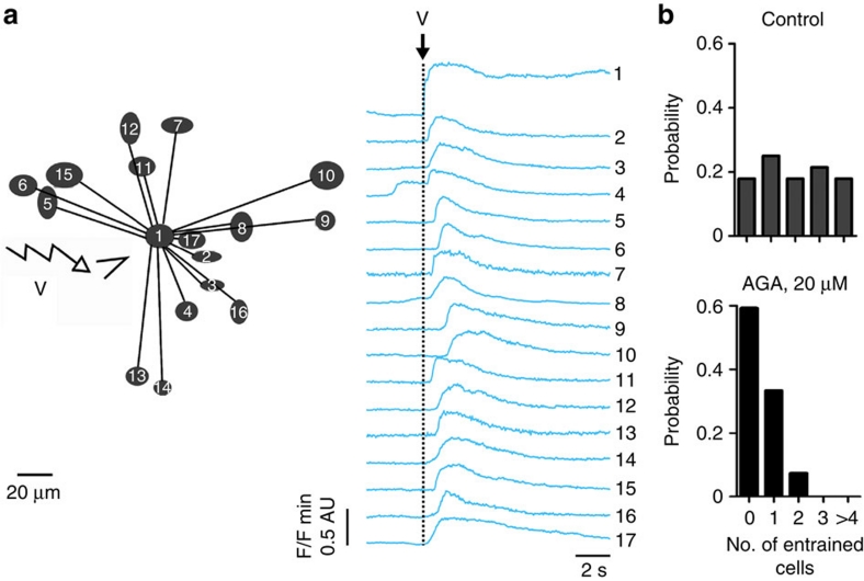
Figure 4. Entrainment of lactotroph activity involves GJ-mediated intercellular communication.
(a) Following application of a 500 ms depolarizing voltage (V) pulse (onset marked with a black dashed line) onto individual DsRed-positive cells within slices from weaned animals, lactotrophs located at distance (left panel) respond with rises in intracellular Ca2+ levels (right panel). The stimulating pipette was touching cell 1 (left panel; triangle) and, for the sake of clarity, only cells that responded with intracellular Ca2+ rises are represented on the connectivity map (AU, arbitrary units). (b) Histograms showing probability of lactotroph entrainment in the absence (top panel; n=28 recordings from five animals) and the presence (bottom panel; n=27 recordings from four animals) of 18-alpha-glycerrhitinic acid (AGA). Note that application of AGA severely impedes cell–cell signal propagation. In all cases, to allow accurate discrimination of entrainment, lactotroph network activity was dampened by constant perfusion with dopamine (100 nM).

