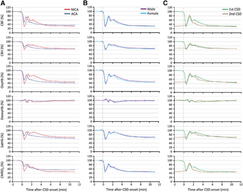
Figure 4.
The hemodynamic and metabolic changes during cortical spreading depression (CSD) in different arterial territories, in male and female mice, and in response to a second consecutive CSD. (A) The hemodynamic and metabolic responses to the first CSD measured within the middle and anterior cerebral artery (MCA and ACA) territories are shown superimposed for direct comparison (n=6 females). Both the magnitude and the duration of hypoperfusion were smaller within the ACA territory. In addition, the second smaller peak observed in MCA territory was virtually absent within the ACA territory. P<0.05 versus MCA in all measured parameters. (B) The hemodynamic and metabolic responses to the first CSD did not differ between male and female mice shown superimposed for direct comparison (n=5 and 6, respectively). Responses were recorded within the MCA territory, and did not differ statistically. (C) The hemodynamic and metabolic responses to two consecutive CSDs are shown superimposed for direct comparison (n=6 females). Responses were recorded within the MCA territory. Note that the ostensibly monophasic response to the second CSD was superimposed on post-CSD oligemia and closely resembled the first CSD with the exception of the initial hypoperfusion. The standard errors of both the magnitudes and the latencies of measured time points are also shown in each panel. Statistical comparisons were not done because of different baselines for the first and second CSDs for all measured parameters.

