
FIG. 1.
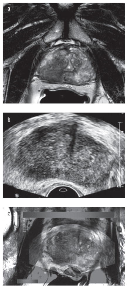
Magnetic resonance imaging-transrectal ultrasonography (MRI–TRUS) fusion in a 67-year-old male with an elevated prostate-specific antigen of 14 ng/dL. (a) Axial T2 weighted magnetic resonance image demonstrates a right-sided low signal intensity area in the right mid-peripheral zone. (b) Transrectal ultrasound image at axial plane shows a hypoechoic lesion at right mid-peripheral zone. (c) Fusion of T2-weighted MRI study with real-time transrectal ultrasound.