
FIG. 4.
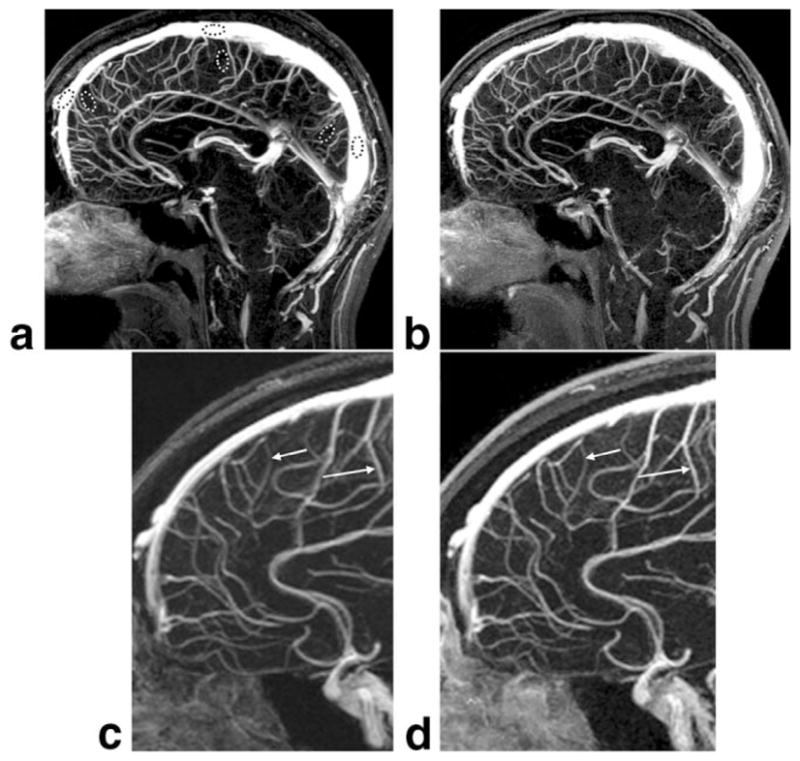
Comparison of midline sagittal partitions taken from reference non-SENSE (a) and R = 4 SENSE (b) whole-brain MR venograms from volunteer 7. Enlargements c and d, which correspond to the dashed box regions of a and b, respectively, illustrate improved vessel sharpness of the SENSE result (arrows, d vs. c) for equal sampling resolution. In this volunteer the SENSE acquisition was done with the second bolus injection. The window and level are normalized to the vessel signal and background.