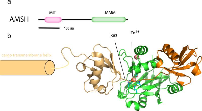
Figure 5.
The zinc isopeptidase AMSH. (a) Domain architecture of AMSH. (b) Structural model for the catalytic complex of AMSH-LP bound to a Lys63-Ub2-modified cargo. The structural model was derived by superimposing the structure of active, zinc-bound, Ub-free form of AMSH-LP (2ZNR) on the structure of the Lys63-Ub2 complex (2ZVN; two shades of orange for the two moieties) with an inactivated mutant lacking the catalytic zinc ion. To illustrate the positioning of the proximal and distal moieties of the Lys63-Ub, the cargo is modeled as a single pass transmembrane protein with the ubiquitin conjugated close to the transmembrane domain. Zinc ions are shown as salmon-colored space-filling spheres.

