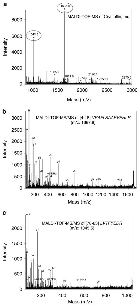
Figure 5.
Mass spectrometry identification of mu crystallin. (a) Peptide mass fingerprint of mu crystallin with peptides marked with ‘*’ matched by MASCOT search against the National Center for Biotechnology Information (NCBI) primate database. The x and y axes show the mass/charge (m/z) ratio and the % abundance of the tryptic peptide fragments, respectively. (b) Matrix-assisted laserdesorption ionization time-of-flight tandem mass spectrometry (MALDI-TOF-MS/MS) analysis of peptide fragment from mu crystallin with m/z of 1667.8 was sequenced as VPAFLSAAEVEEHLR. The corresponding y- and b-series ions and also the immonium ions are shown. (c) A similar MALDI-TOF-MS/MS analysis of peptide fragment with m/z 1042.5 was sequenced as LVTFYEDR.

