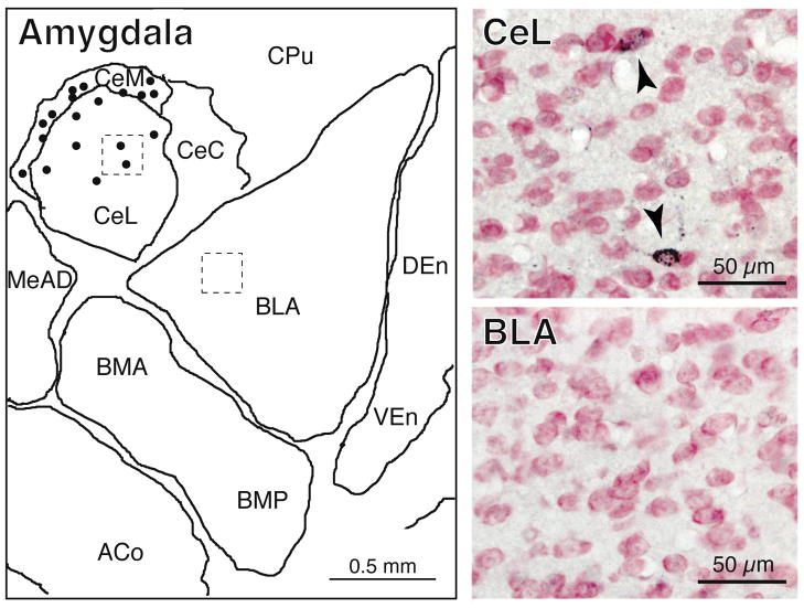
Figure 3.
Schematic and photomicrographs of the amygdala. In the schematic, retrogradely-labeled (CTB+) neurons (dots, left panel) are present exclusively in the central but not the basal, medial or lateral nuclei of the amygdala (left panel). The top and bottom photomicrographs (right panels) correspond to the dashed areas indicated in the central and lateral nuclei of the amygdala, respectively. The arrowheads in the top right panel point to CTB+ neurons. In the bottom photomicrograph, there are no CTB+ neurons. Abbreviations: ACo, anterior cortical nucleus of the amygdala; BLA, basolateral nucleus of the amygdala, anterior part; BMA, basomedial nucleus of the amygdala, anterior part; BMP, basomedial nucleus of the amygdala, posterior part; CeC, central nucleus of the amygdala, capsular division; CeL, central nucleus of the amygdala, lateral division; CeM, central nucleus of the amygdala, medial division; CPu, caudate putamen; CTB, cholera toxin B-subunit; DEn, dorsal endopiriform nucleus; MeAD, medial nucleus of the amygdala, anterodorsal part; VEn, ventral endopiriform nucleus.

