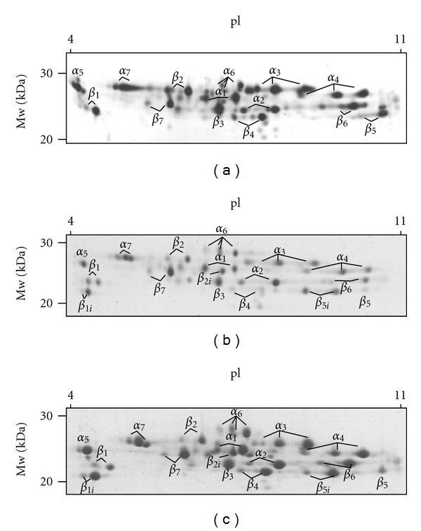
Figure 4.

2D-PAGE of purified 20S proteasomes from (a) red cells (5 μg), (b) BAL-supernatant (20 μg) from ARDS patients, and (c) spleen (30 μg). Detection of protein spots was performed by silver staining and Coomassie BB G250, respectively. Standard 20S proteasome was exclusively detected in red cells (a). Samples of human spleen and of the BAL supernatants from ARDS patients showed both standard and immunoproteasome proteins (panels (b) and (c)).
