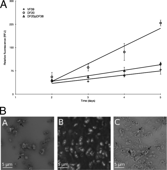
Fig 1.
(A) Accumulation of PHB granules over time. Cells were grown on VMM with mannitol and Nile blue A, and fluorescence was measured after 2, 3, 4, and 5 days of incubation at 30°C. Viable plate counts were used to standardize the fluorescence data. (B) Fluorescence microscopy of PHB granules in cells grown on VMM with mannitol and Nile blue A for 5 days at 30°C. Panel A, wild type; panel B, chvG mutant; panel C, chvG complement.

