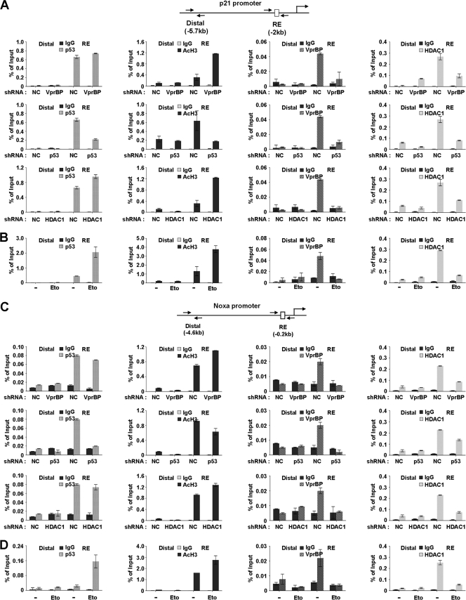
Fig 5.
Dynamics of promoter occupancy of VprBP, p53, and HDAC1. (A) VprBP depletion-promoted H3 acetylation at the p21 promoter. VprBP, p53, and HDAC1 were depleted as for Fig. 4A, and ChIP assays of promoter and distal regions were performed using antibodies specifically recognizing VprBP, p53, HDAC1, and acetyl H3. Input DNA and immunoprecipitated DNA were quantified by qPCR analyses using distal and proximal primer sets. The results are shown as percentages of input, and the error bar indicates the means ± SE. (B) DNA damage-induced dissociation of VprBP. U2OS cells were treated with or without etoposide (100 μM) for 8 h and then analyzed by ChIP analysis of p21 promoter as described for panel A. (C) VprBP depletion-promoted H3 acetylation at the Noxa promoter. ChIP analyses were essentially as described for panel A but over the Noxa gene promoter. (D) DNA damage-induced dissociation of VprBP. U2OS cells were either DMSO treated or etoposide (100 μM) treated for 8 h and then analyzed by ChIP analysis of the Noxa promoter as described for panel B.

