
Fig 1.
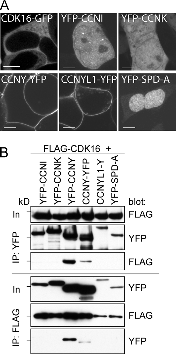
CDK16 interacts with CCNY. (A) 293A cells were transfected for 48 h with expression plasmids for the indicated proteins and imaged on a Leica SP5 confocal microscope using a 63× objective. Bars = 5 μm. (B) 293A cells were transfected for 60 h, and FLAG-tagged CDK16 and YFP-tagged cyclins were immunoprecipitated using anti-FLAG and anti-YFP antibodies and analyzed by immunoblotting. Input (In) is 5% of the protein amount used for IP. The 72-kDa marker is indicated.