
Fig 1.
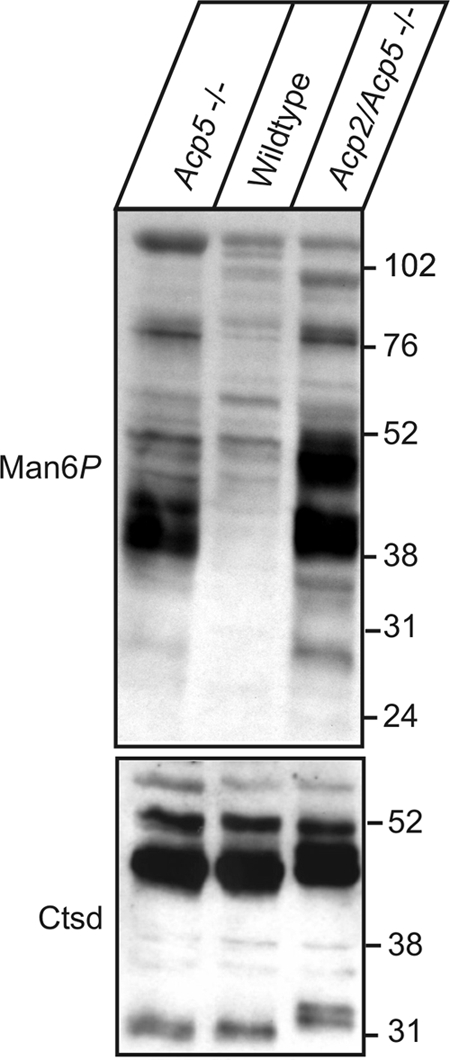
Man6P-containing proteins in liver of Acp5−/− and Acp2/Acp5−/− mice. Eighty micrograms of protein extracts prepared from liver tissue of 8-week-old wild-type, Acp5−/−, and Acp2/Acp5−/− mice was separated by SDS-PAGE and analyzed by anti-Man6P Western blotting (1 μg of scFv M6P-1/ml). The positions of the molecular mass marker proteins (in kilodaltons) are indicated. As a control for equal loading, Western blotting of the lysosomal protein cathepsin D is shown.