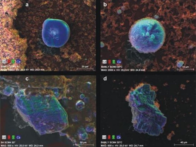
Fig 6.
EDX mapping of bacterial calcite minerals of Arthrobacter sulfonivorans (SCM3) (a and b) and Rhodococcus globerulus (SCM4) (c and d). RT, room temperature; B4, B4 liquid medium (28); B4MLY, modified B4 medium. The SEM images show false colors of red, green, and blue for the local occurrence of the elements carbon, oxygen, and calcium, respectively. A gray level SEM image is underlying each. The shape of the blue-green carbonate minerals depend on the strain; it is round if precipitated in the presence of Arthrobacter sulfonivorans but idiomorphous when formed by Rhodococcus globerulus. Note the differences in magnification and scale bars.

