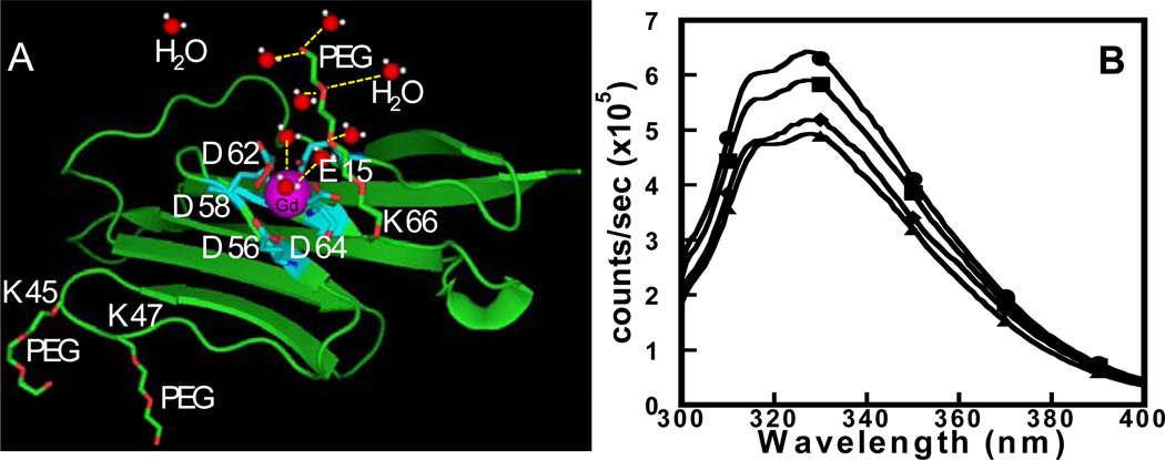
Fig. 1.
(A) Modeled structure of the designed contrast agent ProCA1 is shown with Gd3+ (purple) binding site (cyan), and the cartoon structure of the secondary and outer sphere water molecules (red and white) associated with PEG chain (red and green) are highlighted. (B) The Trp emission fluorescence spectrum of ProCA1-PEG0.6k (●) and ProCA1-PEG2.4k (■) are similar to ProCA1 (◆) and W.T. CD2 (▲), indicating that the PEGylation did not alter the overall conformation of the protein.

