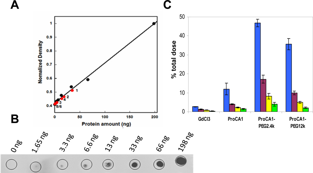
Fig. 5.
(A) PEGylation increased the mouse in vivo blood retention time of the agents monitored by dot-blotting. Appropriate dosages of Gd-ProCA1-PEG2.4k, Gd-ProCA1-PEG0.6k and Gd-ProCA1 were injected intravenously into the mouse via the tail vein. Blood samples (~50 µL) were collected via orbital sinus at different time points. The content of protein MRI contrast agent in the blood was monitored by dot-blotting. Equal volumes of serum were loaded on the plate and identified by the commercial CD2 polyclonal antibody PabCD2. The amount of the ProCA1-PEG2.4k (1) and ProCA1-PEG0.6k (2) left in blood serum were higher than ProCA1 (3) at 3 h. At 19 h, the amount of ProCA1-PEG2.4k (4) left in blood serum is more than ProCA1-PEG0.6k (5) and ProCA1 (6). (B) Results from control samples with different amounts of ProCA1 show that the longer blood retention time was observed for proteins with higher degree of PEGylation and longer PEG chain modifications. (C) γ-radiation of 153Gd isotope. Accumulation of 153Gd in blood circulation (% of total injected dose) for GdCl3, Gd-ProCA1, Gd-ProCA1-PEG2.4k, and Gd-ProCA1-PEG12k at time points of 1 (blue), 4 (purple), 8 (yellow) and 12 h (green) after injection.

