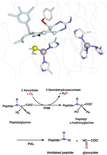
Figure 1.
Top: view of the active site of PHM showing the CuH (right) coordinated to three histidines and the CuM center (left) centers coordinated to two histidines and a methionine. A substrate molecule (di-iodo-YG) is bound in the site close to the M center. (Taken from pdb file 1OPM). Bottom: reactions catalyzed by the PHM and PAL domains of peptidylglycine α-amidating lyase (PAM).

