Figure 4.
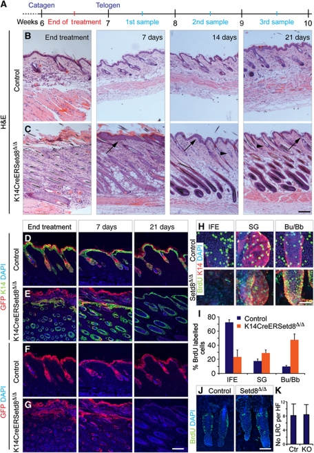
Long-lived progenitors of IFE and sebaceous glands are irreversibly lost in K14CreERSetd8Δ/Δ mice and recovered skin derives from hair follicles (HFs). (A) Schematic overview of the 4-OHT-treatment regime. (B, C) Haematoxylin & Eosin (H&E) staining of skin section from control (B) and K14CreERSetd8Δ/Δ (C) mice at the end of treatment with 4-OHT and after 7, 14 and 21 days of recovery. Arrows point to IFE and arrowheads to sebaceous glands. (D–G) Co-labelling of control (D, F) and K14CreERSetd8Δ/Δ epidermis (E, G) for K14 (green) and GFP (red) (D, E) or GFP (red) only (F, G) after treatment with 4-OHT (end treatment) or after 7 and 21 days of recovery. (H) BrdU incorporation (BrdU; green) and labelling for K14 (red) in the IFE, sebaceous glands (SGs) and HF bulges/bulbs (Bu/Bb) in control (upper panels) and K14CreERSetd8Δ/Δ (Setd8Δ/Δ; lower panels) epidermis. (I) Quantification of BrdU-positive cells from (H). (J) Detection of quiescent label-retaining cells (LRCs) (BrdU; green) in bulges of control and K14CreERSetd8Δ/Δ (Setd8Δ/Δ) mice. (K) Quantification of (J). KO: K14CreERSetd8Δ/Δ. Nuclei are counterstained with DAPI (blue) in (D–H, J). Scale bars: 250 μm (A–G); 50 μm (H) and 75 μm (J).

