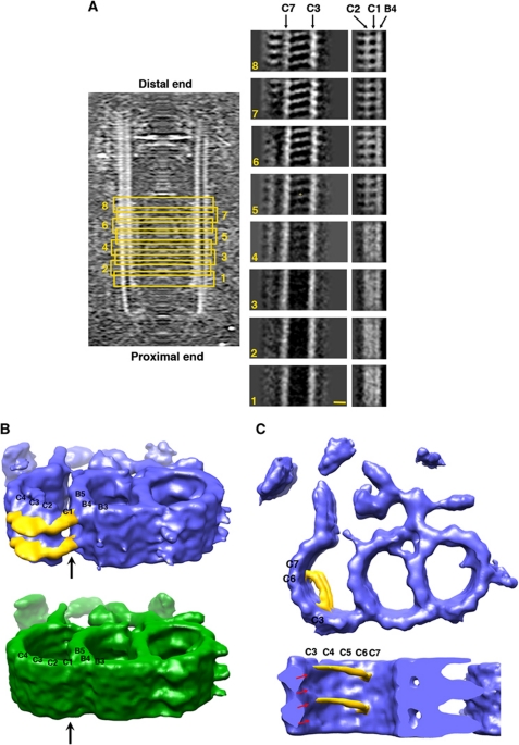
Figure 4.
Longitudinal structural variations observed in the triplets. (A) Subvolumes are divided into eight groups and averaged according to their longitudinal position in the basal body. The relative positions of the group are highlighted with the yellow boxes. Each group contains a triplet segment about 39 nm in length. Structural differences are shown with cross-sectional views of the C-tubule for each averaged triplet, cutting through PFs C3/C7, PFs B4/C1/C2, respectively. The scale bar is 10 nm. (B) Comparison of the two triplet structures resulted from averaging of the proximal half (in green) and the distal half (in blue) separately. The hook-like linkers that connect the B- and C-tubule in the distal half triplet are coloured in yellow. The arrows indicate the positions of C1. (C) Filaments (in yellow) are observed in the lumen of the C-tubule in the distal half of the triplet, with a longitudinal spacing every 8 nm. The tubulin helices are indicated with red arrows. The longitudinal length of the triplets in Figure 4B and C is about 26 nm.

