Figure 1.
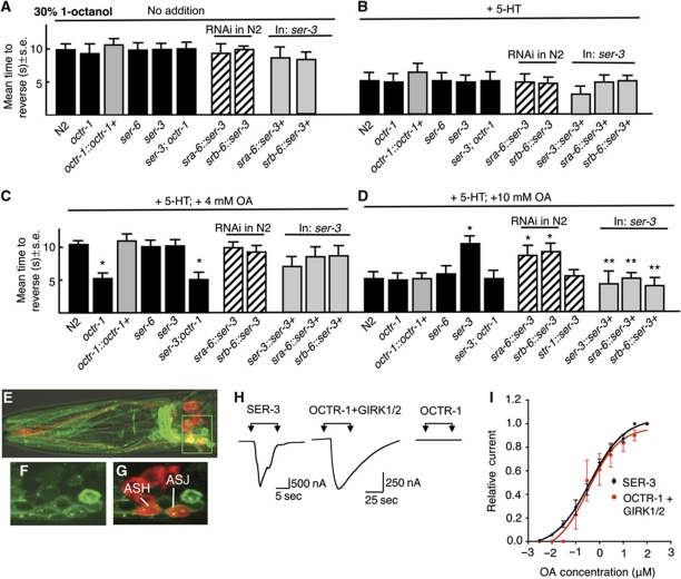
The OA-dependent delay in the 5-HT stimulation of ASH-mediated aversive responses to dilute 1-octanol is modulated by two OA receptors, OCTR-1 and SER-3 in the ASH sensory neurons. (A) Aversive behaviour to 30% 1-octanol was assayed in wild-type, mutant and transgenic animals after incubation in exogenous 5-HT (4 mM) and/or OA (4 or 10 mM), as described in Materials and methods. Black bars, wild-type or null animals; grey bars, null animals expressing a transgene; hatched bars, RNAi in wild-type animals. Data are presented as a mean±s.e. and analysed by two-tailed Student's t-test. *P<0.001, significantly different from wild-type animals under identical test conditions; **P<0.001, significantly different from null mutant. (A) No addition, (B) +4 mM 5-HT, (C) +4 mM 5-HT and 4 mM OA, (D) +4 mM 5-HT and 10 mM OA. (E–G) Fluorescence from ser-3 null animals expressing a ser-3∷ser-3∷gfp translational fusion using confocal microscopy. (E) Merge of GFP fluorescence, (DiD) staining and DIC, (F, G) Z-section from inset in (E) (F, GFP fluorescence; G, Merge of GFP and DiD staining). (H, I) OCTR-1 and SER-3 exhibit nearly identical affinities for OA when heterologously expressed in Xenopus oocytes. (H) OA activates an inward current in oocytes expressing SER-3 or OCTR-1/GIRK1/2, but not in oocytes expressing OCTR-1 alone. (I) OA dose–response curves for oocytes expressing SER-3 or OCTR-1 and GIRK1/2.

