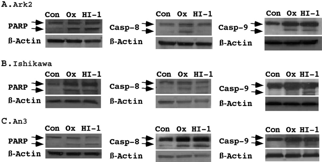
Figure 6.
Western Blot analysis of apoptotic pathways. The cleavage of PARP, caspase-8, and caspase-9, was determined using specific antibodies as described under Materials and methods. The pro-apoptotic proteins and their cleaved products were indicated by arrows on the top and bottom, respectively. For each blot, β-actin levels were measured as a protein loading control. Oxamflatin (0.25 μM) and HDAC-I1 (0.5 μM) treatments resulted in significant activation of PARP in Ark2, Ishikawa and AN3 cells. Oxamflatin displayed preferential effects on caspase-9 cleavage in Ark2 in comparison to Ishikawa and AN3 cells. While oxamflatin induced caspase-8 cleavage in Ark2 cells, HDAC-I1 has little effect in this cell line.

