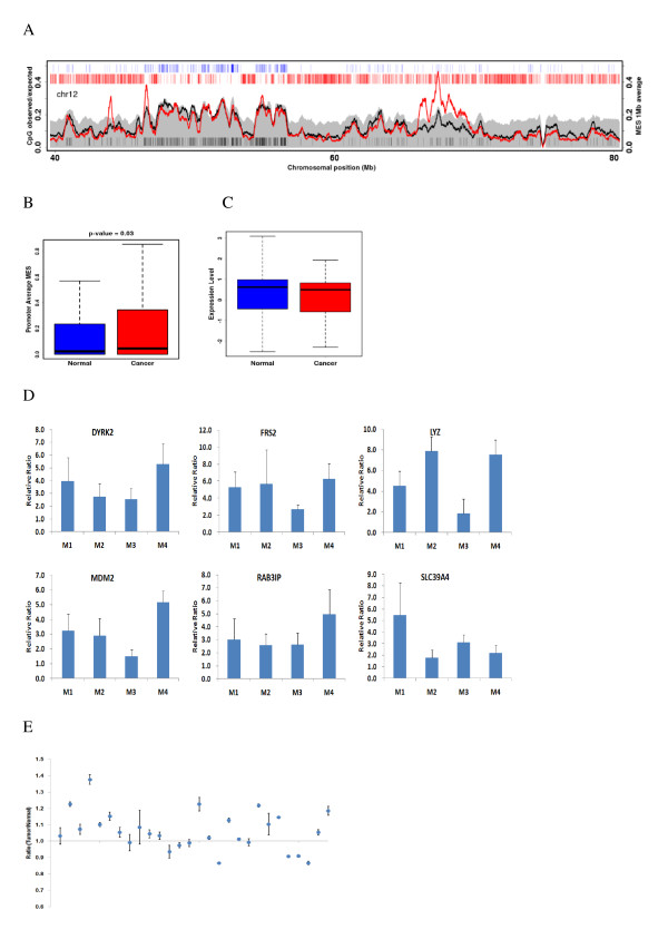
Figure 5.
DNA methylation of LRES regions. (A) Average MES curve for LRES regions in chromosome 12 in normal (black) and cancerous (red) tissue. (B) Average MESs for gene promoters in LRES regions in normal and cancerous tissue. (C) Correlation between gene expression levels and hypermethylation of genes within LRES regions. (D) Methylation enrichment of several selected genes within LRES regions, as assessed by MIRA-qPCR. (E) Methylation ratio at the target site within the upstream region of MDM2, as measured by pyrosequencing.

