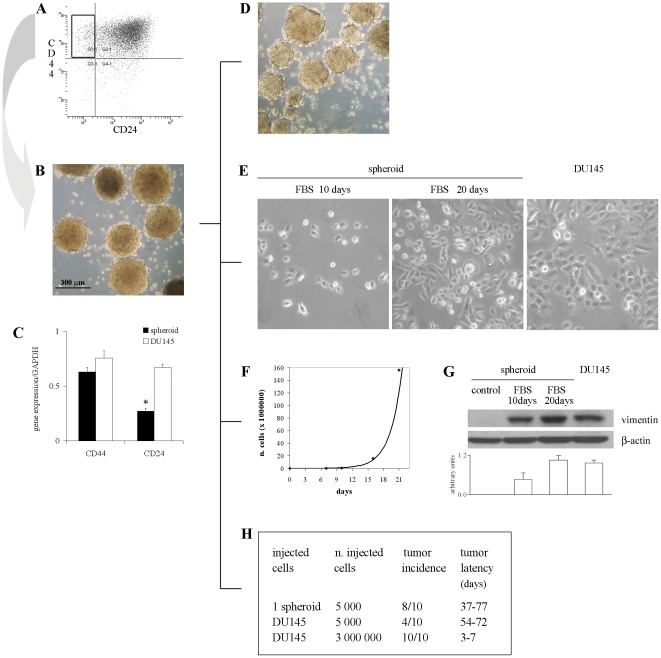
Figure 1. Sorting, culture and characterization of stem-like cells from DU145 cell line.
(A) FACS analysis of CD44 and CD24 expression in DU145 cells. (B) Culture of isolated CD44+CD24− cells growing in SRM as nonadherent prostaspheres (5× objective). (C) Q-PCR of CD44 and CD24 expression in spheroid and DU145 cells. Data are presented as mean ± SEM from 3 experiments. *, p<0.05 vs DU145 cells. (D) Self-renewal capacity of spheroids. Serum supplementation and the withdrawal of growth factors induce the growth of spheroid cells as adherent cells with morphology (E), proliferation rate (F) and expression of vimentin (G) comparable to DU145 cells. Photographs of spheroid cells were taken under a phase contrast microscope after 10 and 20 days of growth in FBS-containing medium (20×). The representative western blots show the expression of vimentin and β-actin in spheroid cells grown in FBS-supplemented medium with respect to control spheroids and DU145 cells. The histogram displays the densitometric quantification of vimentin normalized to β-actin levels. Values are expressed as mean ± SEM from 3 independent experiments. (H) Tumor incidence and latency after injection of 1 spheroid, 5,000 or 3×10⁶ DU145 cells in NOD/SCID mice.

