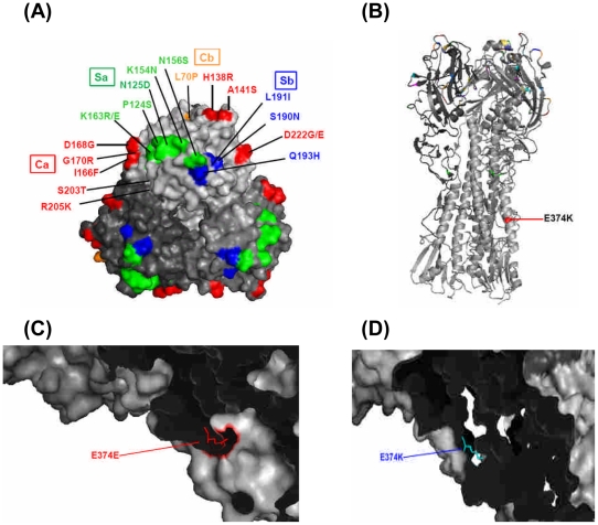
Figure 4. Locations of changed amino acid residues of HA1 and HA2 of pH1N1 virus in Taiwan.
A. Globular head of HA1. Site Ca (Red); Site Cb Orange); Site Sa (Green); Site Sb (Blue). B. Stalk region of HA2. C. Early isolate with E374E residue. D. Late isolate with E374K. All the figures were generated and rendered with the use of MacPyMOL (http://wwwpymolorg). Numbers of amino acids represents the order of the amino acid taking out signal peptide (17 amino acids) and then counting the numbers from initial codon.

