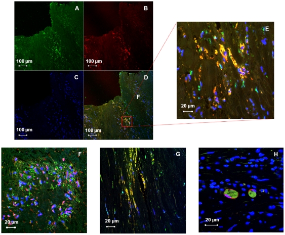
Figure 5. HN is localized to multiple cells of the atheromatous plaque.
Immunofluorescence of the carotid plaque to detect the presence of (A) HN with (B) CD68. (C) Nuclei can be visualized using a DAPI counterstain. (D) Merging of these images demonstrates co-localization of HN with plaque macrophages, (E) seen better with increased magnification. Merged images below demonstrate co-localization of HN with (F) smooth muscle α-actinin, (G) fibroblast vimentin, and (H) dendritic cell fascin.

