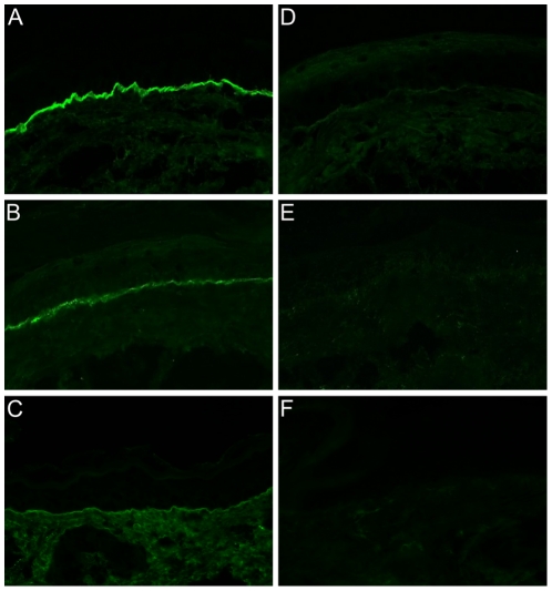
Figure 6. IgG and complement C3 deposition at the basement membrane in experimental bullous pemphigoid.
IF microscopy, performed on frozen sections of a perilesional mouse skin biopsy reveals linear deposition of (A) rabbit IgG, (B) murine C3, and (C) murine IgG at the epidermal basement membrane in a diseased mouse. No deposits of (D) rabbit IgG, (E) murine C3, and (F) murine IgG of a control mouse showing no skin lesions (magnification, ×400).

