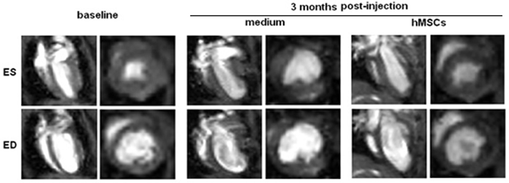
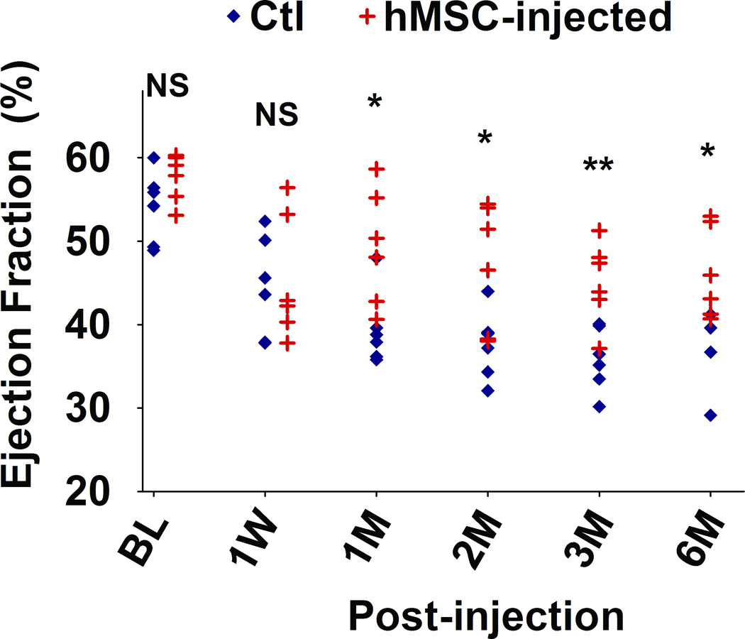
Figure 3. Injection of hMSCs improves cardiac function, as assessed by cardiac MRI.


A. Representative sequential images collected for analysis of the end-systolic (ES) and end-diastolic (ED) volumes from an hMSC-transplanted mouse and a control mouse over a six month period. B. Scatter plot showing left ventricular ejection fraction (LVEF) values of individual mice in hMSC-transplanted versus control groups, collected beginning at one day before injection (baseline, BL), one week (W) and continuing over a six month (M) time period after injection. The mean ± SE was calculated for each group, and repeated-measures ANOVA was used for comparison of LVEFs between control and hMSCs groups (N = 6 mice per group, NS, not significant; **p<0.01; *p<0.05). Beginning at one month after hMSC injection, there was a significant difference in LVEF between groups at each time point.
