
Figure 4. Injection of hMSCs enhances cardiac angiogenesis and attenuates cardiac fibrosis.
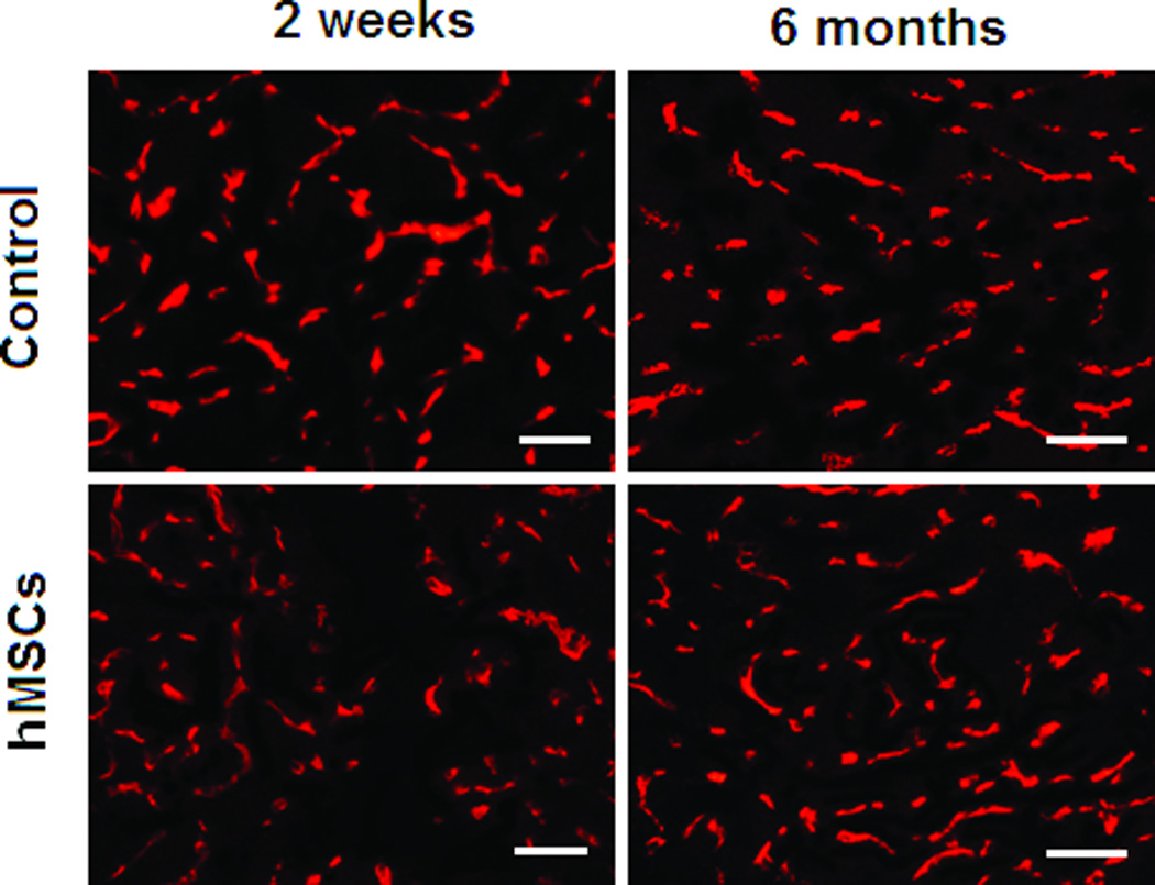
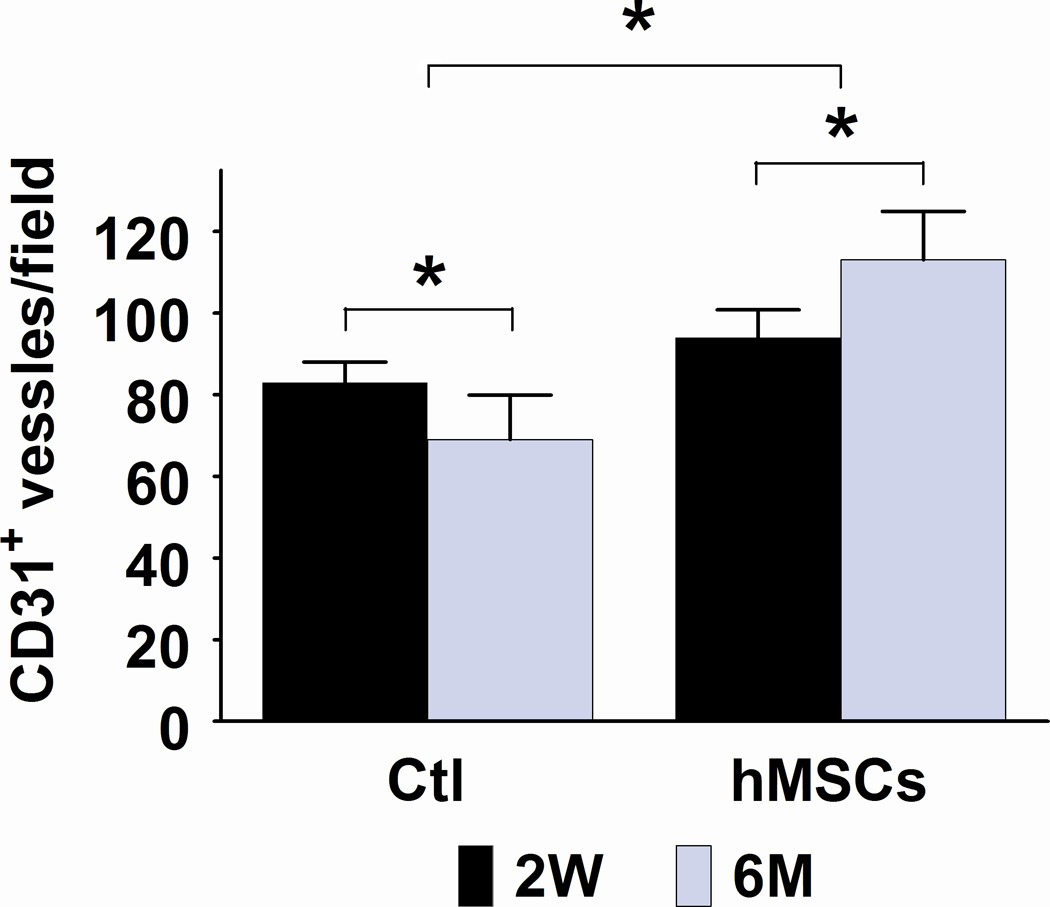
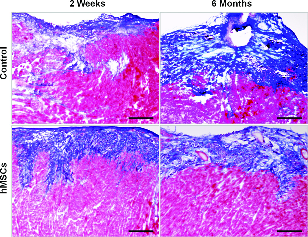
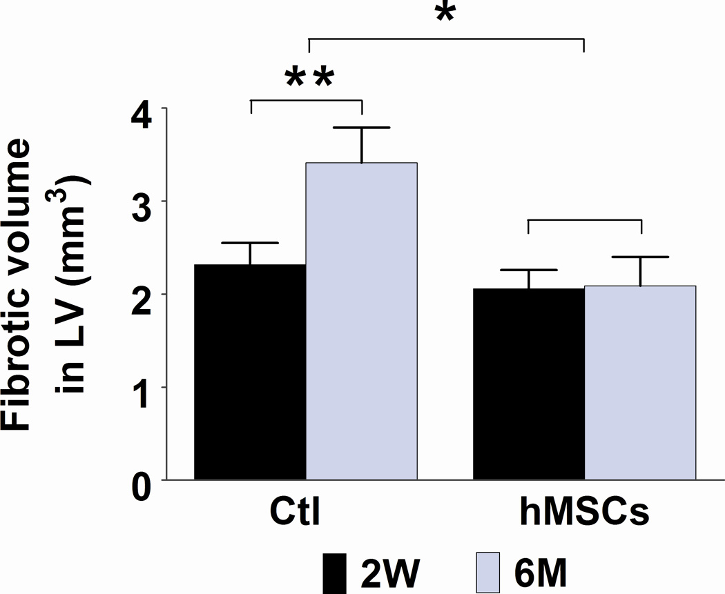
A and C. Representative frozen heart sections from mice two weeks (W) and six months (M) following hMSC or control (medium) injection. Peri-infarct sections were stained with antibody to CD31 to visualize the vessel density (A) and stained with Masson trichrome to evaluate fibrosis (C). Scale bars, 200 µm. B. Quantification of CD31+ cells in peri-infarct sections that are shown in Figure A. The bars represent mean ± standard deviation 16 frozen sections per animal. For each section, the number of CD31+ vessels in 6 fields (400X) was counted and averaged. D. Morphometric analysis of cardiac fibrosis was performed as described the methods and elsewhere.19 The mean ± SE was calculated for each group, and the Student’s t-test was used for evaluate the differences between the groups (N = 3 mice per group, **p<0.01; *p<0.05).