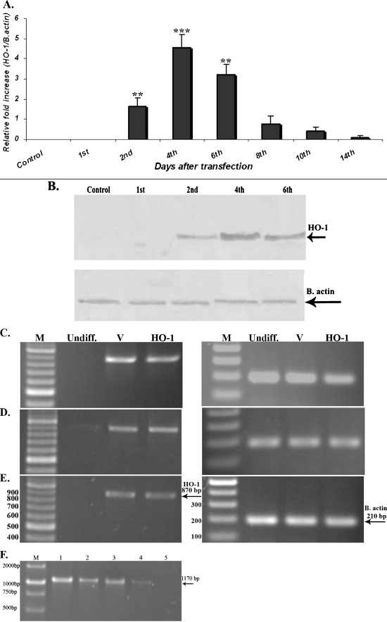
Fig. 1.
Expression of HO-1 gene in MSCs. a Expression of HO-1 mRNA until various time points after infection with adenovirus vectors harboring the HO-1 gene (Ad-hHO-1 data represents mean ± SD; number of replicates = 3, **p < 0.01, ***p < 0.001). b Western blot analysis of the HO-1 protein expression following infection of the MSCs with the recombinant virus. c–e Evaluation of HO-1 expression following MSCs differentiation. HO-1 was expressed in both MSC-V and MSC-HO-1 cells following cell differentiation indicating that HO-1 is expressed in a basal level in these cells, but no expression was detected in the undifferentiated MSCs. c Osteocytes, d adipocytes, e chondrocyte, f extraction of viral DNA and PCR analysis. Following infection of MSCs with the Ad-hHO-1 and induction of osteocyte differentiation, viral DNA was extracted and PCR was performed using pAd/CMV/V5 primers in different time intervals. A strong (1) and a faint band (4) of about 1,170 bp was detected 5 and 12 days post-infection, respectively. However, no bands were observed on day 20. In the two other differentiated cells, adipocytes and chondrocytes, the same results were observed; 2 (8th day), 3 (10th day)

