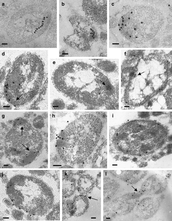
Fig. 5.
Localization of the IbpA protein in A. laidlawii cells. Pattern I: The label was visualized on: some regular structures at the periphery of a cell (a, b), on a cell pole (c), along the structures (probably, cytoskeleton-like) (d), across the structures (e, f). Pattern II: The label was observed at the electron-dense regions of the cell (g, h). Pattern III: The label was present mainly on the membranes (i, j). Pattern IV: The label is predominantly on constrictions of the dividing cells (k, l). Intracellular structures (d–f), electron-dense regions (g), and constrictions (k, l) are pointed by arrows. Bars correspond to 100 nm

