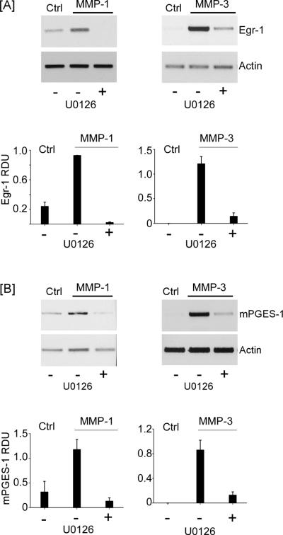
Figure 5.
MMP-induced Egr-1 and mPGES-1 expression are dependent on MAPK kinase. [A] RAW264.7 macrophages (12-well plate; 5 × 10⁵/well) were incubated in DMEM-0.1% LE-BSA (Ctrl) or pre-incubated 30 min in media containing 10 μM U0126 (MAPK kinase inhibitor) followed by the addition of 50 nM MMP-1 or MMP-3, and incubated 1 h. [B] Macrophages (12-well plate; 7 × 10⁵/well) were incubated in DMEM-0.1% LE-BSA or pre-incubated 30 min in media containing 10 μM U0126 followed by the addition of 50 nM MMP-1 or MMP-3, and incubated 18 h. Total RNA was isolated, and mRNA levels for Egr-1, mPGES-1 and actin were determined by PCR. Data are representative blots and the mean levels (± SD) of target mRNAs, presented as RDUs, of 3 experiments.

