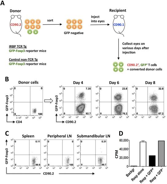
Figure 1. Naïve IRBP-specific T cells convert to FoxP3+ Tregs in the eye.
(A) Experimental paradigm to examine in vivo Treg induction. (B) Progressive conversion of IRBP-specific T cells to FoxP3+ cells in eyes of naïve recipient mice. CD4+GFP–− T cells from IRBP TCR Tg FoxP3-GFP reporter mice on Rag2−/− background were injected into eyes of CD90.1 congenic recipients. On the indicated days after injection, FoxP3 expression in CD4+CD90.2+ cells is shown. Representative experiment of three is shown. (C) Paucity of donor FoxP3+ cells in peripheral lymphoid tissues. Sorted GFP-negative CD4+ T cells from IRBP TCR Tg FoxP3-GFP reporter mice (Rag2−/−) were injected into eyes of CD90.1 congenic recipients. Five and 8 days after injection, cells harvested from the indicated lymphoid organs were stained with CD4-APC and CD90.2-PE. Shown is FoxP3 expression in (7-AAD-excluding) CD4+CD90.2+ donor cells on day 5 after cell injection, when there were approximately 15% converted cells in the eye. Data on day 8 were similar, with 30% converted cells in the eye. Representative experiment of two is shown. (D) In vivo converted cells are functionally suppressive. CD4+FoxP3− T cells isolated from IRBP TCR Tg FoxP3-GFP reporter mice were injected into eyes of CD90.1 congenic recipients. After 8 days CD90.2+FoxP3+ or CD90.2+FoxP3− donor T cells were sorted out and examined in an Ag specific in vitro proliferation inhibition assay against fresh IRBP TCR Tg (Rag2−/−) responder cells. Shown is one of 2 repeat experiments, depicting the proliferation of 50,000 responder cells in presence or absence of the converted or non-converted donor cell populations. Only one well each with donor cells could be set up per experiment.

