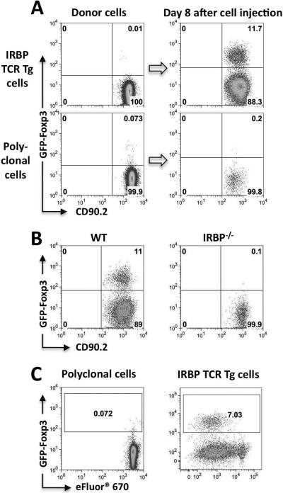
Figure 2. Conversion requires local antigen recognition and is accompanied by proliferation.
(A) Eyes of CD90.1 congenic recipients were injected with CD4+GFP− T cells from IRBP TCR Tg (Rag2−/−) or non-TCR Tg FoxP3-GFP reporter donors, and were analyzed on day 8 after injection. Representative experiment of three is shown. (B) CD4+GFP−T cells sorted from IRBP TCR Tg, Foxp3-GFP reporter mice were injected into eyes of naïve CD90.1 congenic recipients who were either WT (n=4 eyes) or IRBP−/− (n=8 eyes). On day 7 after cell injection, eyes were analyzed for Foxp3 expression on donor cells. Representative experiment of three is shown. (C) Conversion in vivo involves proliferation. Donor cells were labeled with eFluor® 670 and were injected into the eyes of naïve recipients. On day 4, FoxP3 expression and eFluor® 670 dilution in CD4+CD90.2+ was analyzed by flow cytometry. Data are representative of at least three independent experiments.

