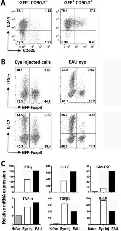
Figure 3. Non–converted cells are primed but are restricted from expressing effector function in the eye.
CD4+GFP–− T cells from naïve IRBP TCR Tg FoxP3-GFP reporter mice (Rag2−/−) were injected into eyes of CD90.1 congenic recipients. (A) Donor cells injected into the eye express activation markers. Seven days after injection, cells from the recipient eyes were analyzed for CD44 vs. CD62L expression on donor cells by flow cytometry. Starting population contained <1% CD44highCD62Llow cells. Shown is a representative one of three independent experiments. (B) Eye-injected donor T cells express low effector function compared to T cells from eyes with actively induced EAU. Seven days after injection, cells isolated from the recipient eyes were pulsed ex vivo with PMA/Ionomycin for 4 hours in the presence of Brefeldin A. Cells stained intracellularly were analyzed for IFN-γ and IL-17 and expression in FoxP3− and in FoxP3+ populations. Cells from eyes of GFP-FoxP3 reporter mice (non-TCR Tg) in which EAU was actively induced by IRBP161–180 immunization were similarly analyzed 7 days after disease onset. Shown are representative plots of four (eye injected) and three (EAU) repetitions. (C) Non-converted GFP− IRBP TCR Tg donor T cells were purified by sorting 8 days after injection into eyes and were analyzed by real-time PCR for expression of pro- and anti-inflammatory cytokines (white columns) compared to CD4+ GFP− cells sorted from eyes of WT mice 8 days after onset of EAU induced by active immunization (black columns). Cytokine mRNA expression is normalized to freshly isolated WT naïve CD4+FoxP3− cells (grey columns).

