
Figure 4. Intracellular signaling in fibroblasts exposed to Gd3+-phosphate.
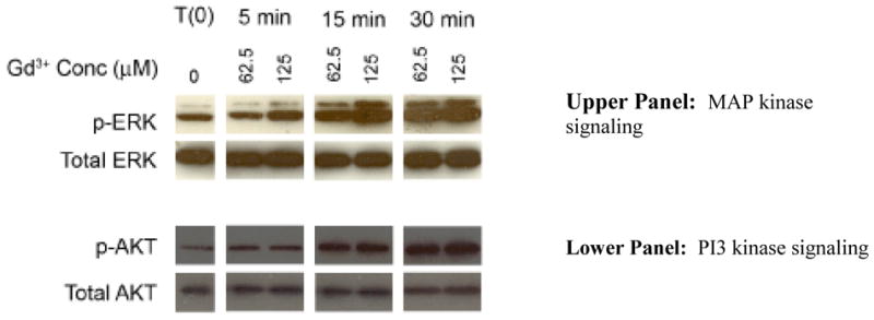
Upper panel: MAP kinase signaling: Cells were treated with Gd3+-phosphate (125 μM Gd3+) for the indicated times. At the end of the incubation period, cell lysates were prepared and assayed for phospho-ERK and total-ERK protein by western blotting. Phospho-ERK and total-ERK from one of three replicate experiments is shown. Lower panel: PI3 kinase signaling. Cells were treated with Gd3+-phosphate (125 μM Gd3+) for the indicated times. At the end of the incubation period, cell lysates were prepared and assayed for phospho-AKT and total AKT protein by western blotting. Phospho-AKT and total-AKT from one of three replicate experiments is shown.