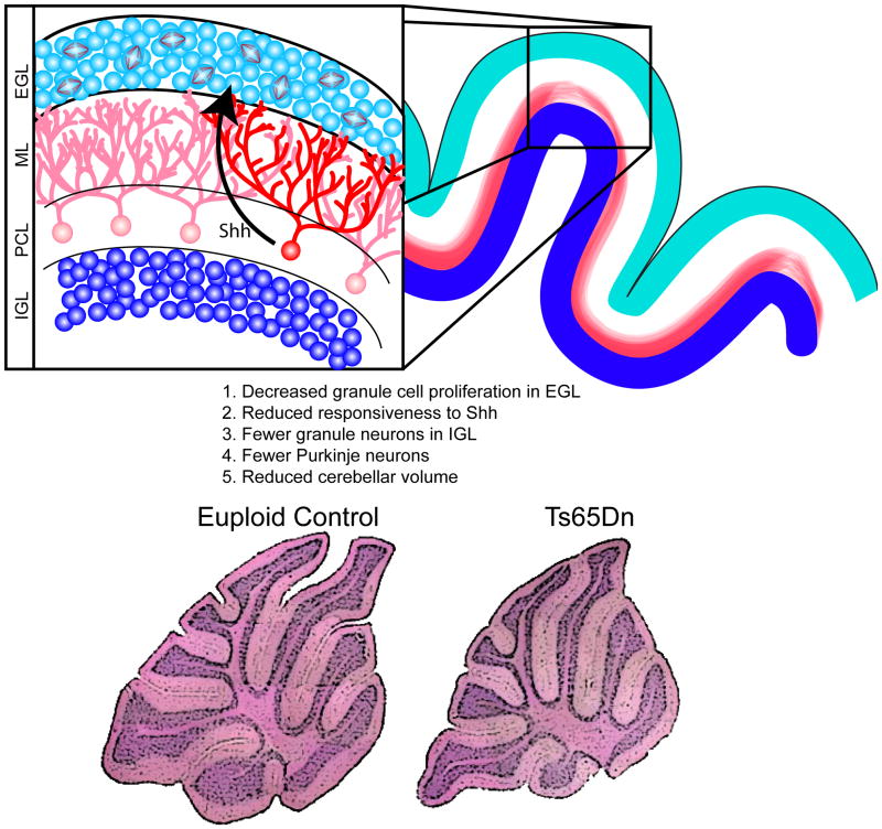
Figure 4. Proliferation deficits in the granule cell precursors (GCPs) lead to reduced cerebellar volume.
Fewer mitotic GPCs are found in the external granule cell layer (EGL) in mouse models of DS [15, 106] and this leads to a paucity of granule cells in the internal granule cell layer (IGL) following their migration inward, past the molecular layer (ML) and Purkinje cell layer (PCL). Reduced granule cell density is seen in the DS cerebellum, as well. The size of the Purkinje cell population is also reduced in trisomic mouse models [15] and, given the reduction in the overall volume of the DS cerebellum [14], this is likely also true in DS. These cell production abnormalities lead to reduced overall cerebellar volume in adult trisomic mice, as shown in the histological sections of euploid (bottom left) and Ts65Dn mouse model (bottom right) [98]. SHH, released by Purkinje cells, is known to influence granule cell production [96, 97], and granule cell precursors in the Ts65Dn cerebellum exhibit reduced responsiveness to SHH compared to euploid; application of a SHH agonist has been used to correct this proliferation defect in the Ts65Dn cerebellum [98]. Histological stainings adapted, with permission, from [98]. Copyright (2006) National Academy of Sciences, USA.

