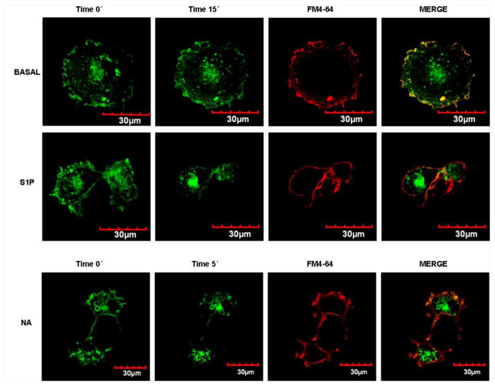
Fig. 10.
Confocal microscopy images of cells expressing α1B-adrenergic receptor-enhanced green fluorescent protein construction. Images were taken at the beginning of incubation (Time 0′) and at the end of the incubation with the reagents (Time 5′ or Time 15′); immediately thereafter, membrane marker FM4-64 (red) was added and images were taken. Merged images (end of incubation-FM4-64) are presented for colocalization (yellow). Cells were incubated in the absence of any agent (BASAL) or presence of 1 μM S1P or 10 μM noradrenaline (NA). Images are representative of 3 to 4 observations using different cell preparations.

