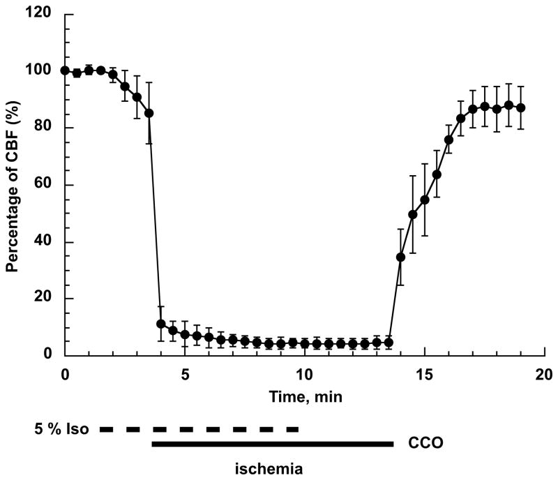
Fig 2.
Changes in cortical blood flow during forebrain ischemia and reperfusion. First minute represents the baseline (100%) blood flow level. Then the isoflurane concentration was increased to 5% and at 3 min the common carotids were occluded (CCO) using microclips. The combination of the high isoflurane level and CCO reduced the blood flow to less than 5% of baseline. At 10 min of CBF recording, the anesthesia was discontinued (0% isoflurane). Following 10 min of CCO the microclips were removed which caused a rapid increase in CBF to about 40% followed by a slower rise. The CBF reached 80 – 90% of baseline at 3 minutes of reperfusion (n=4, error bars represent SD).

