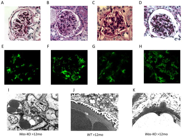
Figure 1. Immune complex deposition and mesangial cell proliferation in WASp deficient mice.
Biopsy specimens from WT controls and Was-KO mice were examined pathologically at different time points. PAS staining of A: WT (>12months) and B: Was-KO (<6months), C: Was-KO (6–12months), D: Was-KO (>12months). Immunofluorescence studies showed immune complex deposition in Was-KO mice. E: IgG, F: IgA, G: IgM, H: C3. Electron microscopic examinations showed mesangial and paramesangial deposits (I), and hump-like deposits (K) in Was-KO mice and no deposits in WT mice (J).

